


 Introduction
Q. What is Excretory System?
Ans: When our cells perform their functions, certain waste products are released into the bloodstream. These are toxic and hence need to be removed from the body. The process of removal of wastes produced in the cells of the living organisms is called Excretory System
Human Excretory System
In humans, the excretory system consists of a pair of kidneys, one pair of ureters, a urinary bladder and a urethra.
Kidney
- Kidneys are reddish brown, bean-shaped structures situated between the levels of last thoracic and third lumbar vertebra close to the dorsal inner wall of the abdominal cavity.
- Each kidney of an adult human measures 10-12 cm in length, 5-7 cm in width, 2-3 cm in thickness with an average weight of 120-¬170g.
- Towards the center of the inner concave surface of the kidney is a notch called hilum through which ureter, blood vessels, and nerves enter.
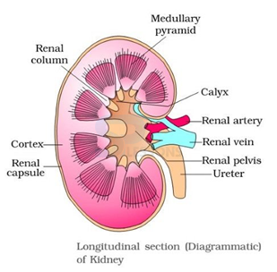
Image: Longitudinal Section of kidney
Source: NCERT Text Books
- Inner to the hilum is a broad funnel shaped space called the renal pelvis with projections called calyces.
- Inside the kidney, there are two zones, an outer cortex, and an inner medulla. The medulla is divided into a few conical masses (medullary pyramids) projecting into the calyces (singularity: calyx).
- Each kidney has nearly one million complex tubular structures called nephrons, which are the functional units.
- Each nephron has two parts - the glomerulus and the renal tubule.
- Glomerulus is a tuft of capillaries formed by the afferent arteriole - a fine branch of renal artery. Blood from the glomerulus is carried away by an efferent arteriole.
- The renal tubule begins with a double-walled cup-like structure called Bowman’s capsule, which encloses the glomerulus.
- The tubule continues further to form a highly coiled network - proximal convoluted tubule (PCT).
- A hairpin shaped Henle’s loop is the next part of the tubule which has a descending and an ascending limb.
- The ascending limb continues as another highly coiled tubular region called distal convoluted tubule (DCT).
- The DCTs of many nephrons open into a straight tube called collecting duct, many of which converge and open into the renal pelvis through medullary pyramids in the calyces.
- The Malpighian corpuscle, PCT, and DCT of the nephron are situated in the cortical region of the kidney whereas the loop of Henle dips into the medulla.
- In a majority of nephrons, the loop of Henle is too short and extends only very little into the medulla. Such nephrons are called cortical nephrons.
- In some of the nephrons, the loop of Henle is very long and runs deep into the medulla. These nephrons are called juxta medullary nephrons.
- The efferent arteriole emerging from the glomerulus forms a fine capillary network around the renal tubule called the peritubular capillaries.
- A minute vessel of this network runs parallel to the Henle’s loop forming a ‘U’ shaped vasa recta. Vasa recta is absent or highly reduced in cortical nephrons.
Biology - Related Information
General Science - Biology - Excretory System
General Science - Biology - Deoxyribonucleic Acid
Human Reproductive System
Respiratory System
Human Reproductive System
