


 Introduction
Q. What is Neural Control and Coordination?
Ans: The functions of the organs/organ systems in our body must be coordinated to maintain homeostasis. Coordination is the process through which two or more organs interact and complement the functions of one another. The neural system provides an organized network of point-to-point connections for a quick coordination. The endocrine system provides chemical integration through hormones.
Neural System
The human neural system is divided into two parts:
- 1. The peripheral neural system (PNS)
2. The central neural system (CNS)
- The CNS includes the brain and the spinal cord and is the site of information processing and control.
- The PNS comprises of all the nerves of the body associated with the CNS (brain and spinal cord).
- 1. Afferent fibres → tissues/organs to brain.
2. Efferent fibres → brain to tissues/organs.
- The afferent nerve fibres transmit impulses from tissues/organs to the CNS and the efferent fibres transmit regulatory impulses from the CNS to the concerned peripheral tissues/organs.
- The PNS is divided into two divisions called somatic neural system and autonomic neural system.
- The somatic neural system relays impulses from the CNS to skeletal muscles while the autonomic neural system transmits impulses from the CNS to the involuntary organs and smooth muscles of the body. 1. Somatic Neural System → Brain to Voluntary muscles. 2. Autonomic Neural System → Brain to Involuntary muscles.
- The autonomic neural system is further classified into sympathetic neural system and parasympathetic neural system.
- The brain is the central information processing organ of our body, and acts as the ‘command and control system’.
- It controls the voluntary movements, balance of the body, functioning of vital involuntary organs (e.g., lungs, heart, kidneys, etc.), thermoregulation, hunger and thirst, circadian (24-hour) rhythms of our body, activities of several endocrine glands and human behavior.
- It is also the site for processing of vision, hearing, speech, memory, intelligence, emotions and thoughts.
- The human brain is well protected by the skull. Inside the skull, the brain is covered by cranial meninges consisting of an outer layer called dura mater, a very thin middle layer called arachnoid and an inner layer (which is in contact with the brain tissue) called pia mater.
- The brain can be divided into three major parts: (i) forebrain, (ii) midbrain, and (iii) hindbrain.
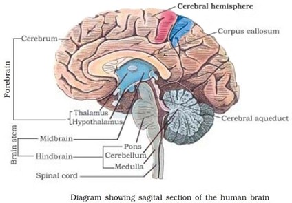
Image: Sagital Section of human brain
Source: NCERT Text Books
- The forebrain consists of cerebrum, thalamus, and hypothalamus.
- Cerebrum forms the major part of the human brain. A deep cleft divides the cerebrum longitudinally into two halves, which are termed as the left and right cerebral hemispheres.
- The hemispheres are connected by a tract of nerve fibres called corpus callosum.
- The layer of cells which covers the cerebral hemisphere is called cerebral cortex. The cerebral cortex is referred to as the grey matter due to its greyish appearance. The neuron cell bodies are concentrated here giving the colour.
- The cerebral cortex contains motor areas, sensory areas, and large regions that are neither clearly sensory nor motor in function. These regions called as the association areas are responsible for complex functions like intersensory associations, memory, and communication.
- Fibres of the tracts are covered with the myelin sheath, which constitute the inner part of cerebral hemisphere. They give an opaque white appearance to the layer and, hence, is called the white matter.
- The cerebrum wraps around a structure called thalamus, which is a major coordinating centre for sensory and motor signaling.
- Another very important part of the brain called hypothalamus lies at the base of the thalamus. The hypothalamus contains a number of centres which control body temperature, urge for eating and drinking. It also contains several groups of neurosecretory cells, which secrete hormones called hypothalamic hormones.
- The inner parts of cerebral hemispheres and a group of associated deep structures like amygdala, hippocampus, etc., form a complex structure called the limbic lobe or limbic system. Along with the hypothalamus, it is involved in the regulation of sexual behaviour, expression of emotional reactions (e.g., excitement, pleasure, rage and fear), and motivation.
- The midbrain is located between the thalamus/hypothalamus of the forebrain and pons of the hindbrain. A canal called the cerebral aqueduct passess through the midbrain.
- The dorsal portion of the midbrain consists mainly of four round swellings (lobes) called corpora quadrigemina. Midbrain and hindbrain form the brain stem.
- The hindbrain comprises pons, cerebellum, and medulla (also called the medulla oblongata).
- Pons consists of fibre tracts that interconnect different regions of the brain.
- Cerebellum has very convoluted surface in order to provide the additional space for many more neurons.
- The medulla of the brain is connected to the spinal cord. The medulla contains centres which control respiration, cardiovascular reflexes, and gastric secretions.
- You must have experienced a sudden withdrawal of a body part which comes in contact with objects that are extremely hot, cold pointed or animals that are scary or poisonous.
- The entire process of response to a peripheral nervous stimulation, that occurs involuntarily, i.e., without conscious effort or thought and requires the involvement of a part of the central nervous system is called a reflex action.
- The reflex pathway comprises at least one afferent neuron (receptor) and one efferent (effector or excitor) neuron appropriately arranged in a series.
- The afferent neuron receives signal from a sensory organ and transmits the impulse via a dorsal nerve root into the CNS (at the level of spinal cord). The efferent nueuron then carries signals from CNS to the effector. The stimulus and response thus forms a reflex arc as shown below in the knee jerk reflex.
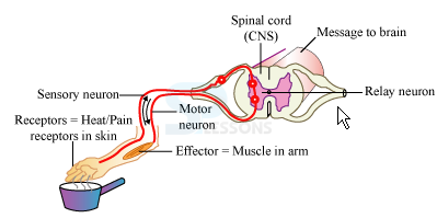
Image: Diagram of a Reflex Arc
Source: NCERT Text Books
Biology - Related Information
General Science Facts
General Science Practice Set
Important General Science Quiz
General Science - Biology - DNA RNA
